
What You Need to Know About CT Angiography
When it comes to heart and vascular health, fast and accurate diagnosis is of vital importance. CT angiography is a valuable diagnostic method offered by modern medicine at this point. Compared to traditional angiography, it is preferred for critical diagnoses, especially in emergency situations, due to its non-invasive nature and fast process that can be completed in just 15-20 minutes.
What is CT Angiography?
CT angiography is the process of imaging the blood vessels in our body using a computed tomography (CT) scanner. Also known as CT angiography, this method provides a detailed examination of the internal structure of blood vessels. It plays a significant role in the early diagnosis of heart diseases. It is also referred to as virtual angiography, 3D computerized heart angiography, non-surgical heart angiography, or heart tomography.
In particular, coronary CT angiography allows for the three-dimensional examination of the coronary arteries that supply the heart. Additionally, the aorta, main neck arteries, lung arteries, liver, kidney, and brain arteries can also be visualized using this method. Unlike traditional angiography, CT angiography allows for the evaluation of not only the inner part of the artery but also its outer part and the structure of the arterial wall. The procedure is performed within an average breath-holding time of 10-15 seconds. This examination is completed much more quickly than traditional angiography. The patient can return to their normal life immediately after the procedure.
CT angiography can be used to detect vascular occlusions, aneurysms, plaque buildup (calcifications), vascular ruptures, narrowings, and blockages. However, if a narrowing is detected, conventional angiography may be required to determine the treatment method. Thanks to the development of multi-slice computed tomography technology in recent years, CT angiography can now be performed much faster and with higher resolution images.
When is it Used?
CT angiography plays a critical role in the early diagnosis of various vascular problems. It is particularly preferred in patients with complaints such as heart disease, chest pain, and shortness of breath. It is very useful for risk assessment in individuals with a family history of heart disease, who smoke, and who have high blood pressure or cholesterol problems.
Beyond the question of what CT angiography is, we can list the following situations in terms of its areas of use:
- Evaluation of suspected coronary artery disease and risk
- Detection of pulmonary embolism
- Emergency situations requiring immediate intervention, such as aortic aneurysm and aortic dissection
- Narrowing or aneurysms in the brain arteries
- Carotid artery narrowing in the neck
- Graft control after bypass surgery
- Follow-up of patients with stents
- Causes of heart muscle diseases and genetic heart rhythm disorders
It is the first choice diagnostic method in cases of acute chest pain because it provides quick results. It is also used as a preventive approach in patients who are at risk of heart attack but do not yet show serious symptoms.
In addition, CT angiography offers a safe alternative in elderly patients who cannot undergo invasive angiography, individuals at risk of bleeding, or those with unsuitable vascular structures. However, it is not suitable for individuals whose heart rate cannot be lowered, those with contrast medium allergies, or patients with renal failure.
In cardiology, it is preferred in patients with unclear stress test results or in situations where conventional catheter angiography is inadequate and risky. It also provides effective results in the evaluation of congenital heart diseases and heart valve problems. For this reason, CT angiography is increasingly used in the evaluation of vascular health. It has also become a reliable and rapid diagnostic method.
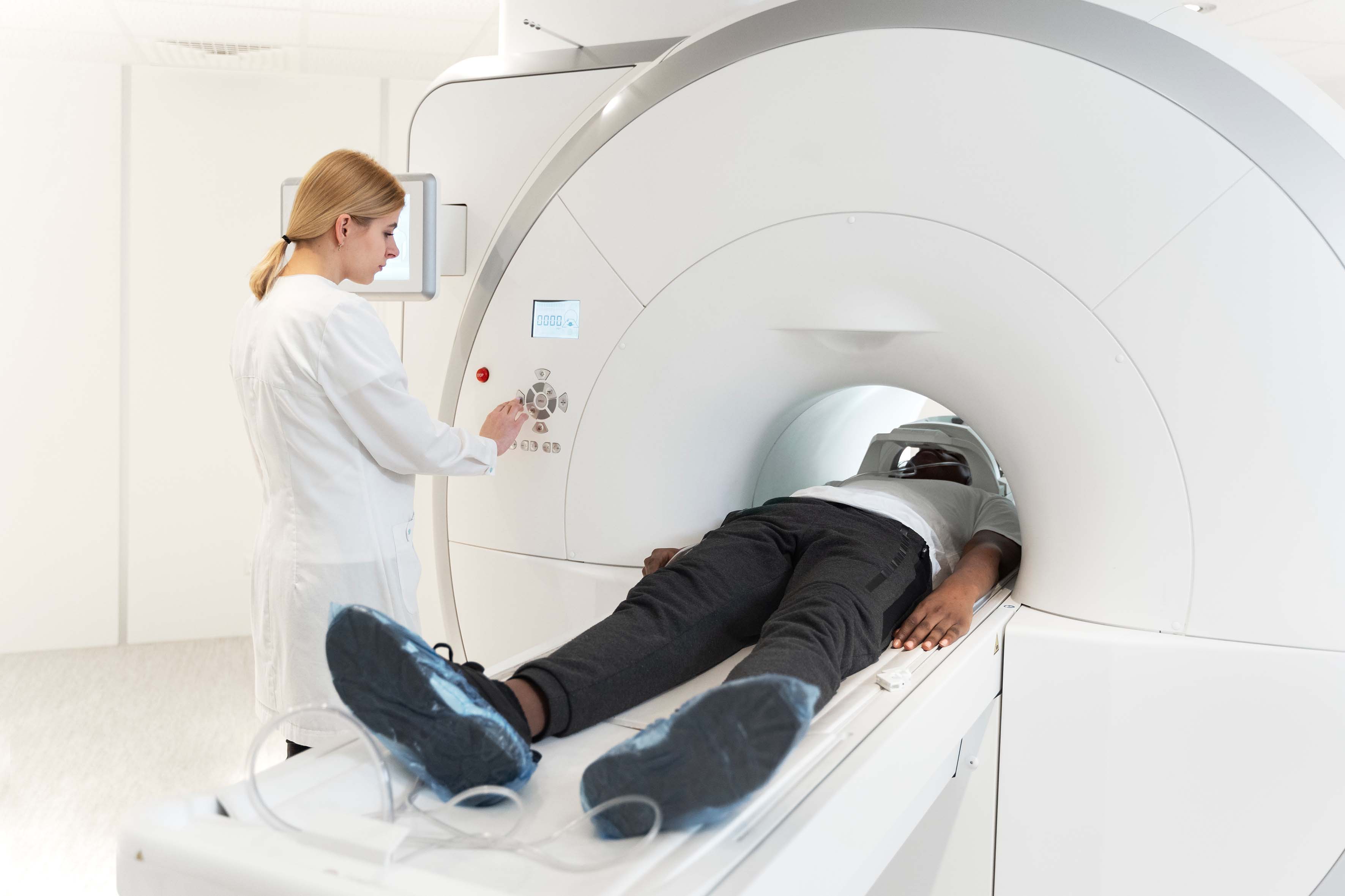
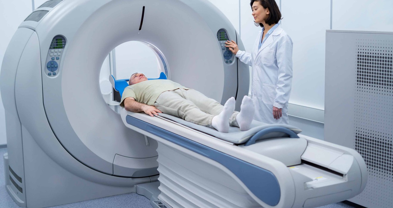
How is CT Angiography Performed, and What Should Be Considered?
Pre-procedure preparation is a critical step that directly affects the success of CT angiography. You should avoid eating anything and only drink water for approximately 4-6 hours before the procedure. Additionally, you should avoid caffeinated beverages such as tea, coffee, and energy drinks for 12 hours before the procedure. This is because these beverages can increase heart rate, making it difficult to obtain clear images.
On the day of the procedure, you must first inform your doctor of any allergies you may have and any medications you regularly take. It is of vital importance to know whether you have an allergy to contrast agents. In addition, a blood test (creatinine) is performed to assess your kidney function. During CT angiography, an iodine-containing contrast agent is used to obtain clearer images of your blood vessels.
The CT angiography procedure is performed in the following stages:
- Opening of the blood vessel and administration of medication to lower the heart rate if necessary (the ideal heart rate should be around 60 beats per minute).
- Positioning you in the appropriate position for the tomography device.
- Informing you about breathing commands and practicing them.
- Injection of contrast material into the arm vein.
- Imaging of the target area within seconds.
During the procedure, you may experience a sensation of warmth in your body, a metallic taste in your mouth, and a feeling of urinary incontinence due to the contrast agent. The procedure is completed in approximately 15-20 minutes, including the active imaging time and follow-up time.
After the procedure, it is recommended that you drink plenty of water to help eliminate the contrast agent from your body. Generally, after a short observation period (15-30 minutes), you can return to your normal daily activities. This method, which is also used to examine vital arteries such as the aorta, provides rapid diagnosis, especially in cases of suspected aortic dissection. There are no special restrictions other than avoiding heavy work and lifting.
Advantages, Risks, and Results of CT Angiography
CT angiography is one of the most valuable tools in modern imaging technology. It significantly facilitates the diagnostic process with the benefits it offers. The greatest advantage of this method is that it is non-invasive, meaning it does not require surgical intervention. Unlike traditional angiography, it can be performed without inserting a catheter into the groin, so the risk of complications is quite low.
Other notable advantages of CT angiography include:
- It can be completed in a short time, typically 5-15 minutes.
- It provides high-resolution and 3D images.
- There is no pain during or after the procedure.
- It can evaluate vital structures such as the brain, neck, lungs, and aorta, in addition to the heart vessels.
- It can measure the structure of plaques on the arterial walls and the amount of calcium present.
- It can be used for follow-up in patients who have undergone stent placement or bypass surgery.
However, as with any medical procedure, there are some risks associated with CT angiography that should be considered. First, an allergic reaction to the contrast agent used during the procedure may occur. Additionally, although rare, there is a risk of cancer due to exposure to radiation. In patients with kidney failure, the contrast agent may further impair kidney function.
CT angiography should be avoided during pregnancy. Breastfeeding mothers should not breastfeed their babies for 24-48 hours after the procedure. However, despite all these risks, the accuracy rate of the procedure is quite high.
CT angiography results are life-saving in the rapid detection of conditions requiring urgent intervention, such as aortic dissection. By providing detailed information about the degree of vascular narrowing, plaque characteristics, and coronary anatomy, it enables doctors to determine the correct treatment strategy. With all these features, CT angiography has become an indispensable diagnostic tool in modern cardiology.